_(2).jpg)
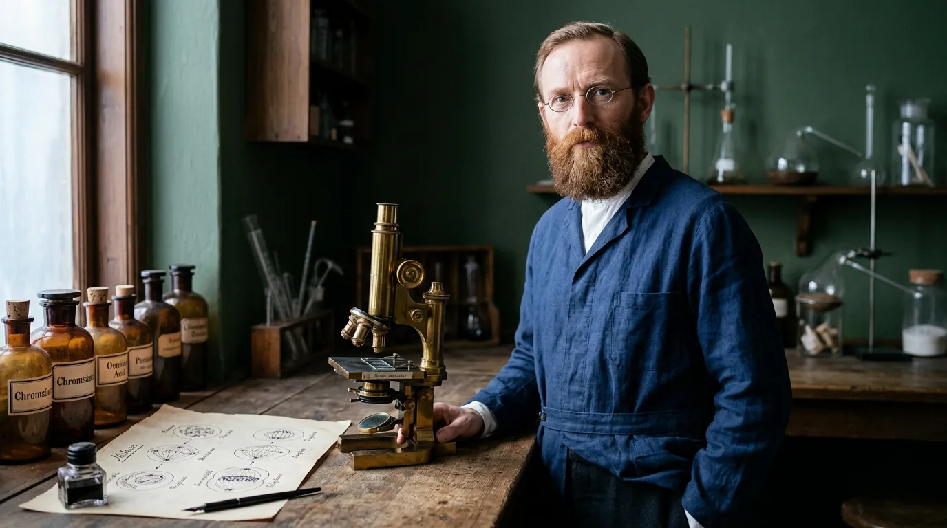
Walther Flemming
1843–1905 · Cell division, chromatin and mitosis
Walther Flemming traced the movement of stained nuclear material as cells divided, giving biology a clearer description of mitosis and chromatin.
Why is this scientist famous?
Walther Flemming used stains and microscopes to reveal how nuclear material changes during cell division, laying foundations for modern mitosis research.
Mitosis remains vital to teaching growth, tissue repair and how dividing cells pass chromosome copies to daughter cells.
Walther's story
Looking closely at changing cells
Walther Flemming was born near Schwerin, in the German state of Mecklenburg, on 21 April 1843. He studied medicine at several German universities and completed his medical doctorate at Rostock in 1868. After working in research and medical posts, he became a professor in Prague in 1873. In 1876 he took up a professorship of anatomy at the University of Kiel. There, he used microscopes to investigate changes inside animal cells.
Why staining mattered
Not every structure inside a cell is easy to see through an ordinary microscope. Flemming used chemical stains to make nuclear material stand out more clearly. He called the strongly staining material chromatin, from a word referring to colour. He compared fixed, stained tissue with observations of living cells, including cells from salamanders. This combination helped him notice patterns that earlier observers had struggled to follow.
The orderly movement of chromosomes
Flemming observed threads of nuclear material change shape as a cell approached division. He followed how the threads condensed, lined up and separated as two new nuclei formed. His detailed drawings revealed that this was a sequence of events, not a sudden mysterious change. He helped establish the term mitosis for this type of nuclear division. The word chromosome came later; Flemming did not know the molecular structure of DNA.
Pictures that transformed biology
In 1882 Flemming published Cell Substance, Nucleus and Cell Division, bringing his findings together. His drawings enabled other scientists to compare the stages of division and investigate their meaning. He also contributed improvements to chemical fixation and staining of tissues. Other scientists developed the modern understanding of chromosomes, genes and inheritance much later. Flemming’s evidence provided a crucial early view of the physical process of cell division.
A foundation for studying growth
Flemming continued investigating cells and their nuclei during his years at Kiel. Illness forced him to retire from his professorship in 1901. He died in Kiel on 4 August 1905. Today, students can use microscopes or images to recognise stages of mitosis much as Flemming sought to do. His work helped bridge the idea that cells divide with evidence of how material inside them behaves.
What did they discover?
Describing mitosis
He documented changing nuclear structures through a sequence of stages as animal cells divided.
Chromatin
He named the nuclear material that stained strongly under the microscope, helping researchers follow its changes.
Microscope staining and fixation
His careful combination of living observations and chemically prepared tissues made cell division easier to investigate.
Cell division drawings
His 1882 book assembled illustrations that helped other scientists build on his research into chromosomes and nuclei.
Amazing facts
He completed medical qualifications at the University of Rostock in 1868.
He became a professor in Prague before joining the University of Kiel in 1876.
Salamander tissues were among the living materials he used to observe dividing cells.
The word chromatin refers to nuclear material that takes up microscope stains.
His observations distinguished strongly stained chromatin from more lightly stained nuclear material.
His important book Cell Substance, Nucleus and Cell Division appeared in 1882.
He used both observations of living cells and preparations preserved and stained with chemicals.
He left his Kiel professorship because of ill health in 1901.
Timeline
Born near Schwerin, in Mecklenburg.
Completed his medical doctorate at the University of Rostock.
Became professor of histology and embryology in Prague.
Appointed professor of anatomy at the University of Kiel.
Published pioneering studies of nuclear changes during cell division.
Published Cell Substance, Nucleus and Cell Division.
Retired from his Kiel professorship owing to ill health.
Died in Kiel.
Related Super Scientists
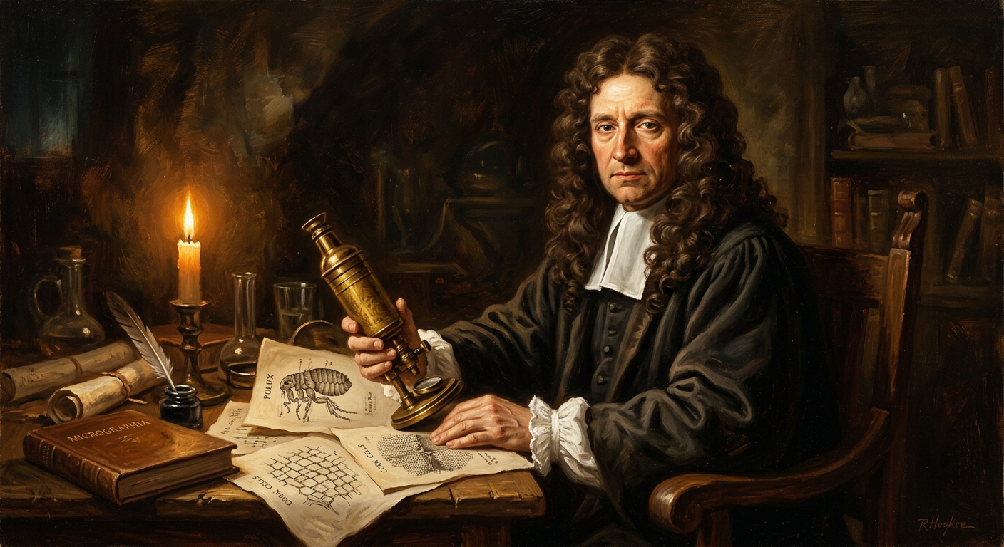
Robert Hooke
1635–1703 · Biologist and Physicist
Coined the word "cell" after examining cork under a microscope and formulated Hooke’s Law on springs.
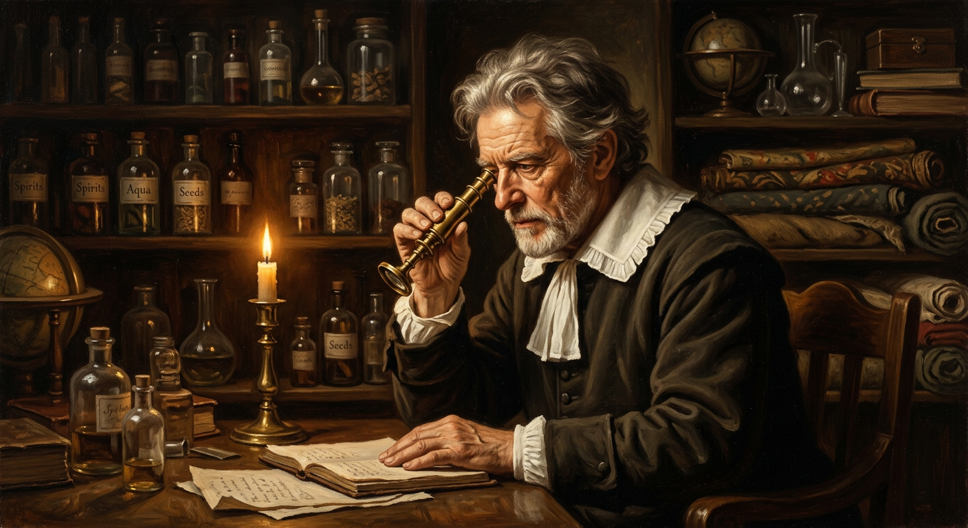
Antonie van Leeuwenhoek
1632–1723 · Biologist
First person to see and describe microorganisms — tiny living creatures invisible to the naked eye.